
2025 CiteScore: 0.1

The Journal is now indexed by Scopus.

Vol 10, No 3 (Summer 2023)
Introduction: The SARS-CoV-2 virus is a group of viruses that cause disease in birds and
mammals and currently has more than 398.572.320 infected and 5.770.578 victims worldwide.
Common clinical symptoms are fever, headache, dyspnea, sore throat, dry cough, abdominal pain,
diarrhea, and nausea. On the other hand, the results of various studies related to oral and maxil-
lofacial manifestations in patients with COVID-19 have reported contradictory results in different
places, which shows the role of different geographical and individual factors in the occurrence of
oral manifestations. The aim of the present study was to review the oral and maxillofacial manifes-
tations in patients with or improved COVID-19 infection.
Materials and Methods: This study is a narrative review. In this study, research published
in Pubmed, Google Scholar and Scopus databases has been reviewed by a review method and with
a keyword search strategy.
Results: 4011 articles were found from the mentioned databases, and after deleting unrelated
researches (3607 cases) and duplicate researches (358 cases), 46 articles remained for final review.
Then, from this number, those research that were presented in the scientific conference and were
in the form of abstracts or did not have a correct statistical population were excluded from the
study (10 cases).
Conclusion: COVID-19 is a new and rapidly evolving disease, and many of its features, in-
cluding its associated oral manifestations, are still under investigation. No agreement has yet been
reached on the nature and classification of oral lesions in COVID-19 and their causal relationship
to disease progression. This limitation is reflected in systematic reviews such as the present article.
In addition, there is a significant shortage of high-quality studies on the prevalence of these oral
manifestations and this limitation affects the prevalence estimates presented in this study.
Keywords: Oral manifestations; Maxillofacial manifestations; Sars covid_19.
Introduction: Odontogenic lesions related to impacted teeth are common findings in routine
dental exams, with the potential to cause tooth pain, displacement, bone expansion, or even bone
erosion. Thus, their early detection is important. This study aimed to evaluate the frequency of such
lesions based on patient age, gender, and location at the Tehran University of Medical Sciences,
School of Dentistry, over five years.
Materials and Methods: This retrospective study evaluated records from the oral pathology
department, covering a five-year period. The association of odontogenic lesions with impacted
teeth was assessed. Data on lesion type, patient age and gender, involved jaw, lesion location, and affected
teeth were extracted from patient records. Statistical analysis was performed using SPSS 24.
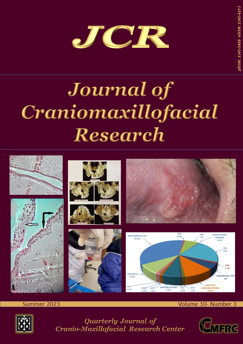
Results: Out of 815 odontogenic lesions, 44.4% were associated with impacted teeth. The patient
distribution was 52.48% male and 47.52% female. Lesions were most prevalent in the third decade
of life (24.58%), and third molars (59.39%) were frequently involved. The posterior mandible
(68.5%) and mandibular third molar (55%) were common sites. Dentigerous cysts had the highest
frequency (35.4%), with odontogenic cysts (developmental) occurring more frequently (70.7%)
than odontogenic tumors.
Conclusion: The study underscores a significant association between odontogenic lesions and
impacted or unerupted teeth. Periodic follow-ups are crucial for retaining impacted teeth based
on these findings.
Keywords: Tooth; Impacted; Odontogenic cysts; Odontogenic tumors; Jaw; Pathology; Oral.
Introduction: Oral Squamous Cell Carcinoma is the 6th most prevalent cancer worldwide.
The global increase in frequency and mortality of oral SCC has been shown. There are few studies
about the risk factors for oral SCC in Iran. This study is based on finding a possible relationship
between oral SCC and some lifestyle factors in the Iranian population.
Materials and Methods: This case-control study was conducted at the Iran Cancer Institute
in Tehran. The controls were matched to the cases by age and gender and socio-economic status.
A number of 204 individuals (102 cases and 102 controls) were interviewed by using a structured
questionnaire to obtain data regarding oral hygiene, dietary factors, smoking habits, alcohol consumption
and lifestyle risk factors for oral squamous cell carcinoma. Results were analyzed by chisquare
and Fisher exact test. P values less than 0.05 were considered significant.
Results: The mean age of cases at diagnosis was 60.03 years and 52.9% were male. The analysis
showed that poor oral hygiene, low intake of fruits and vegetables (P<0.001), low intake of dairy
(P=0.029), alcohol consumption (P=0.015), cigarette smoking (P=0.002) in years and pack per year
of smoking (P<0.001) are independent risk factors for oral SCC.
Conclusion: The study provided strong evidence that poor oral hygiene, poor dietary factors,
smoking habits and alcohol consumption play an etiological role for oral SCC in the Iranian population.
Keywords: Risk factors; Oral squamous cell carcinoma.
Introduction: Recurrent aphthous stomatitis (RAS) is a common ulcerative inflammatory
condition of the oral mucosa. Considering the contradictory findings of previous research about
the association between anxiety and RAS, the present study aimed to determine the prevalence of
RAS and its association with anxiety among dentistry students.
Materials and Methods: In this descriptive-analytical study, a total of 241 dentistry students
of G. University were divided into two groups (those with a history of RAS and healthy
subjects) after recording their demographic and required information. Also, Cattell’s questionnaire
was used to evaluate the level of anxiety. The results were analyzed using SPSS v.21 software, and a
p-value of <0.05 was considered statistically significant.
Results: 2.40% of subjects suffered from RAS. This condition was more prevalent in winter, and
its incidence was significantly different between the two groups in terms of the presence of a family
history of RAS (p=0.01), food allergy (p=0.05), and regular exercise (p=0.040). Subjects with RAS
had a significantly higher anxiety intensity in the areas of lack of ego strength, superego susceptibility,
and ergic tension compared to subjects without RAS (p<0.0001).
Conclusion: The results showed that some areas of anxiety, including lack of ego strength, can
be predictors of RAS. Therefore, teaching life skills such as anxiety management and its timely
diagnosis and treatment can play an essential role in the prevention and treatment of RAS.
Keywords: Recurrent aphthous stomatitis; Anxiety; Dentistry students.
Introduction: Dentistry is not only an evolving science but also an art with good eye-hand
coordination. A variety of magnification devices are used in dentistry to increase the clarity and
accuracy of examinations and surgeries. The purpose of this study is to evaluate knowledge, attitude
and practice toward using dental magnification among dental residents and dental professors.
Materials and Methods: In this cross-sectional descriptive study, all professors and residents
were studied by census. A questionnaire was administered to the clinical faculty to assess
three parameters: knowledge, attitudes and practice of magnification devices among dental residents
and masters. The questionnaire consisted of 4 parts: demographic information, knowledge
(11 questions), attitude (7 questions), and practice (4 questions). Data were analyzed by SPSS 23
statistical software and analyzed by T-tests and one-way analysis of variance (ANOVA) and Chi-
Square.
Results: A total of 81 dentists completed the research questionnaire, of which 50 (61.7%) were
male and 31 (38.3%) were female. Most dentists answered knowledge questions about depth and
field of view changes with increasing magnification and the ability to adjust eye convergence under
a microscope (74.1%). Also, the results of ANOVA statistical test did not show a significant difference
between the scores of dentists’ attitudes according to the field of specialization.
Conclusion: Although the skill of dentists was acceptable at using magnifying devices, they had
little knowledge about the importance of this issue. The most important reasons were mentioned
lack of access and high cost of using these devices.
Keywords: Attitude; Dentistry; Knowledge; Magnifying devices; Practice.
Dentigerous cyst is one of the most common odontogenic cysts of the jaw that slowly grows around
an unerupted tooth crown. Nasopalatine duct cyst is a non-odontogenic common jaw cyst. Co-occurrence
of nasopalatine duct cyst and dentigerous cyst in the same location of the same jaw is extremely
rare. Odontogenic and non-odontogenic cysts occurring simultaneously in non-syndromic
patients is rare. The lesions must be fully extracted by surgery. We report the treatment procedure
of a patient suffering from this rare phenomenon.
Keywords: Odontogenic cyst; Nonodontogenic cyst; Impacted tooth.
A 39-year-old male was referred to the Oral and Maxillofacial Medicine department of Alborz
University of Medical Sciences Dental School with a lesion that he had noticed for 3 weeks. The
patient had no signs or symptoms and no cigarette and alcohol consumption. Because the lesion
was placed at the lateral posterior portion of the tongue which is a common place for oral malignancies
a biopsy was done. The pathology result was Squamous cell carcinoma grade 2. The patient
was referred to Imam Khomeini Cancer Institute for further treatment.
Keywords: Oral scc; Oral cancer; Squamous cell carcinoma.

 |
All the work in this journal are licensed under a Creative Commons Attribution-NonCommercial 4.0 International License. |