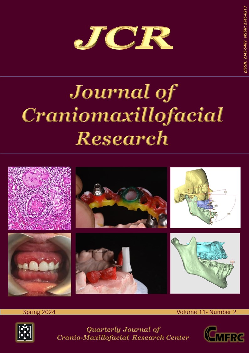
The Journal is now indexed by Scopus.
Vol 11, No 2 (Spring 2024)
Original Article(s)
-
Introduction: Scientists have developed solutions, for oral health by leveraging advancements in medicine. These solutions, known as “ reconstructive dentistry “ aim to address the limitations and short lifespan of restorative methods. Unlike restorative treatments, reconstructive dentistry focuses on not only restoring the structure of tissues but also their physiological functions. To achieve this, it requires an effort among dentists, biologists, stem cell researchers, material scientists, tissue engineers and other experts. Consequently, this study was carried out to create and evaluate a fellowship program called “Reconstructive Medicine in Dentistry.” The program aims to foster an approach towards reconstruction, in dentistry. Materials and Methods: This research involved three stages. Utilized a mixed method approach. In the first stage, we developed a design based on the Kern model, which involved conducting a review of relevant studies to extract and determine the goals and topics related to medical reconstruction, in dentistry. Expert panel meetings were held to finalize these goals and topics. For each target group, we identified the learners, learning environment and educational strategies. We also made arrangements for implementing the program. Established evaluation methods for both students and the program itself. Subsequently, we implemented the designed curriculum for two groups of students. Finally, we evaluated the program’s effectiveness through questionnaires and semi structured interviews, with students, professors and organizers. The collected data was analyzed using statistics. Results: In 2020 and 2021, two groups of students were meticulously selected from disciplines such as maxillofacial surgeons, oral medicine specialists, periodontists, endodontists, and more, after passing the entrance exam. The primary objective of this program is to educate graduates with the skills to restore missing tissues in the mouth, jaw and facial regions using cutting edge regenerative medicine techniques. The curriculum was developed in collaboration with experts from areas like tissue engineering, dental specialization (including maxillofacial surgeons) biomaterials and developmental biology. Both students and professors expressed satisfaction, with the program. Conclusion: A group of professors, from specialties came together to implement a fellowship program in reconstructive dentistry. The main goal of this program was to train specialists in tissue reconstruction for patients who have suffered jaw and facial injuries; by using advanced methods in medicine. Additionally, this initiative can also be a step towards strengthening the university’s movement towards third and fourth-generation universities. The findings from this study can provide insights, for those involved in planning and implementing interdisciplinary fellowship programs. Keywords: Dentistry; Regenerative medicine; Curriculum design; Interdisciplinary.
-
Introduction: Medication-related osteonecrosis of the jaw (MRONJ) is an important uncommon complication. Due to its complexity, its prevention requires a multidisciplinary approach, involving physicians and dental clinicians. Materials and Methods: This study aimed to assess the knowledge level of physicians prescribing bisphosphonates in Tehran, Iran, about dental considerations in such patients and the prevention and treatment of MRONJ in 2019. This descriptive, cross-sectional study evaluated 100 physicians (rheumatologists, endocrinologists, oncologists, and orthopedists) practicing in Tehran. A questionnaire comprising a demographic section and knowledge questions regarding dental considerations in patients taking bisphosphonates was administered among the physicians. The frequency of qualitative variables such as gender, type of specialty, and physicians’ responses to each question was calculated, and the knowledge scores were analyzed separately based on the physicians’ specialty types using one-way ANOVA followed by Tukey’s test for pairwise comparisons. The effect of different variables on knowledge scores was analyzed by simple regression. Results: The mean knowledge score of physicians was 5.19±1.78 (range 2-8). The mean knowledge score of oncologists was significantly higher than that of endocrinologists (5.88 versus 4.52, P=0.03). No other significant differences were noted. Work experience (P=0.04), age (P=0.02), orthopedics specialty (P=0.05), and oncology specialty (P=0.006) had significant effects on the knowledge score. Conclusion: Considering acquiring about 50% of the total score, physicians seem to have limited knowledge about dental considerations in patients taking bisphosphonates. Keywords: Knowledge; Tehran; Bisphosphonates; Bisphosphonate-associated osteonecrosis of the jaw.
-
Introduction: Mucormycosis, an infection, with a high death rate, requires understanding its symptoms, diagnosis methods and treatment options due, to its growing occurrence. The research employs a molecular method to evaluate the diagnosis of the mucormycosis. Materials and Methods: In this study, we obtained 30 samples from patients undergoing diagnosis and conducted DNA extraction. Additionally, DNA extraction was carried out on 30 tissue samples suspected of infection in paraffin blocks. Subsequently, PCR and Real-time PCR were performed using targeted primers, for mucormycosis followed by analysis of the results. Results: In the research findings among 30 liquid samples 8 tested positive, for mucor using the PCR method. 10 using the Real-time PCR method. Similarly, out of 30 tissue samples, 9 cases showed mucor presence with the PCR method and 11 cases, with the Realtime PCR method. Conclusion: In this study, real-time PCR and PCR techniques showed promising and faster results in detecting Mucor than the culture approach. The molecular methods provided results that could be of great use for scenarios. Keywords: Mucormycosis; Diagnosis; Real-time PCR.
-
Introduction: To evaluate undergraduate students’ clinical ability to extract teeth, we created a new, coordinated, and quantitative assessment form containing nine items that were required to measure the various skills, using the visual analog scale. Materials and Methods: A pilot study was performed with 30 students, each of whom was rated by three examiners. In addition, 118 students (59 fourth-year and 59 fifth-year) were reviewed halfway through the year and at their final examinations. The assessment form was then used to evaluate students’ abilities for tooth extraction throughout the academic year 2022–2023. Results: High inter-examiner reliability and a significant association of mean scores (p<0.001) between three examiners at the beginning and final of the block for both 4th and 5th students. Both groups showed considerable improvement in their mean scores between the beginning and final examinations. The result shows the association between socio-demographic characteristics of patients treated by fourth and fifth-stage students, (52.54% and 54.24%) of the participants were males in fourth and fifth-stage students respectively. At the same time (47.46% and 45.76%) of the participants were females in fourth and fifth stage students respectively. The age of majority of the participants was more than 30 years old, representing (76.27%), and only (10.17%) were between 25-30 years in fourth stage students, and (8.47%) were between 25-30 years in fifth stage students. Conclusion: The use of a newly developed assessment scale during tooth extraction offered an objective, standardized, and feasible method for the assessment of clinical skills of undergraduate students for both formative and summative purposes. Keywords: Dental extraction; Clinical performance; Undergraduate dental students.
-
Introduction: Orthodontic treatment interferes with oral hygiene and can discoloration. Therefore, given the importance of tooth discoloration for patients, the present study determined the effect of orthodontic treatment on tooth color changes. Materials and Methods: Thirty-six patients under orthodontic treatment were evaluated. Photographs were taken before and after orthodontic treatment to evaluate color changes of the teeth. The photos were evaluated by experts and laypeople. The researcher completed a questionnaire consisting of each patient’s demographic data and treatment information (the composite resin type, the bonding agent type, and the bracket and wire types). Then, the relationships between these variables and tooth discoloration were analyzed. McNermar test was used to compare tooth color distribution status before and after orthodontic treatment. Chi-squared or Fisher’s tests were used to compare the tooth discoloration distribution status in terms of each variable studied. Spearman’s correlation coefficient was used to determine the relationship between demographic variables and tooth discoloration. Statistical significance was set at P<0.05. Results: Tooth colors were different before and after treatment. There were no significant relationships between tooth discoloration and the variables of composite resin type, bonding agent, bracket type, and gender. However, the relationship between age and tooth discoloration was significant. Conclusion: Various factors affect tooth color changes after orthodontic treatment. Although the majority of the factors evaluated in the present study did not alone have a significant relationship with tooth discoloration, it can be claimed that tooth discoloration due to orthodontic treatment is a multifactorial finding with several confounding factors. Keywords: Discoloration; Orthodontic treatment; Tooth color.
-
Introduction: Improvement of students’ academic performance is the main goal of educational centers, because the academic performance of individuals is the basis of their success at every juncture. This study aimed to investigate the relationship between study habits and academic achievement motivation in Qazvin dental students in 2022-2023. Materials and Methods: This study was a descriptive-analytical study that was performed on all dental students of Qazvin University of Medical Sciences. Data collection tool was three questionnaires, the first part contained demographic and background information including age, sex, marital status, semester and second part contained the PSSHI study habits questionnaire which was developed by Palesani and Sharma and included 45 questions. The third part contains Hermens questionnaire to measure the motivation of academic achievement. The validity of the questionnaires was obtained using experts’ opinions and its reliability was obtained using Cronbach’s alpha test. The collected data were entered into SPSS software version 26, then using descriptive statistics (mean, standard deviation, etc.) and statistical analysis including independent t-test and ANOVA were analyzed. Results: The results of this study showed that the educational motivation of most students of Qazvin dental school was high which had no significant relationship with sex, age, marital status and term (p>0.05). There was a significant relationship between academic motivation and study habits of students (p˂0.05). Conclusion: The results of this study showed that the study habits of Qazvin dental students are moderately desirable, so it seems that students are not familiar enough with learning facilities and study skills. Therefore to enhance the study skills of students, teaching Workshops will be included in the curriculum. Keywords: Study; Habits; Academic; Motivation; Dentistry.
-
Introduction: Posterior impaction of the maxilla leads to spontaneous rotation of the mandible and these rotations are often accompanied by soft tissue and skeletal changes. The present research aims to determine the effects of posterior impaction of the maxilla on mandible’s Autorotation in patients with anterior open bite.Materials and Methods: Using a 3D reconstructed model of 25 patients with anterior open bites, this descriptive study is conducted. The construction model of the posterior segment of the mandible design was subjected to 2, 3, 5, and 7mm posterior impaction of the maxilla around the ANS axis without any mandibular intervention, using the available CT scan and ProPlan CMF software. Following this, the autorotation and anterior open bite correction were assessed. A basic linear regression test was used to examine the effects of various variables on the anterior open bite closure at various impaction rates. Results: The rise, in impact rate led to an increase in the byte closure rate. With 2, 3, 5 and 7mm posterior impaction of the maxilla, the bite closure was not significantly affected by maxilla length, mandible length, U1-SN angle, ANS-PNS angle with the maxillary occlusal plane, or mandibular incisor angle with the mandibular plane. Nevertheless, during the 5mm posterior maxillary impaction procedure, there was a 0.2mm increase in the open bite closure for every 1 degree increase in IMPA; this number is statistically significant. (p<0.001). Conclusion: The amount of bite closure increased along with the posterior impaction of the maxilla. All other variables did not significantly affect bite closure rate, with the exception of the IMPA variable in 5mm impactions. Keywords: Maxillary posterior impaction; Mandible autorotation; Anterior open bite; ProPlan CMF software.
Case Report(s)
-
This case report describes the rehabilitation of the Esthetic-Zone of the maxillary jaw of a 70-year-old female patient by using 4 implants (BEGO Semados® RS/RSX) with the challenging position of her implants due to her age and channel access of the Fixtures and also the Difficulty in supplying desired prosthetic parts due to existing problems. In the beginning, the treatment plan was to reconstruct the mouth using conventional restoration with UCLA abutments, but due to the unavailability of non-hex abutments As well as the failure to achieve the desired Esthetic demands of the patient by using this treatment plan, especially in the anterior implant area of the jaw, it Innovatively was decided to use a combination of cylindrical abutments (BEGO MultiPlus system) and UCLA abutments to Design and fabrication of the Final restoration. Keywords: Dental implant; Dental prosthesis; Static zone; Internal and external connection; Challenging case.
-
Crohn’s disease (CD) is a process of gastrointestinal mucosa inflammation that could be seen in any part of the gastrointestinal tract from oral to anus. Oral lesions could be the first sign in most patients. The lesions in Crohn’s disease are divided to specific and nonspecific lesions. The specific lesions are less common than nonspecific ones. Tag-like lesions and cobblestones are some of the specific lesions and ulcer is a non-specific lesion. The diagnosis of Crohn’s disease is based on a combination of symptoms, enteroscopy, gastroscopy, capsule endoscopy, histopathology and imaging. Oral involvement of CD is also known as oral Crohn’s disease (OCD). Early diagnosis of OCD may be the most influential factor in controlling it. This case report presents a 47-year-old female patient with gingivitis and gingival erythema and enlargement as her chief complaint which was the first sign of CD. Keywords: Pathology; Oral; Crohn’s disease; Gingival enlargement.


