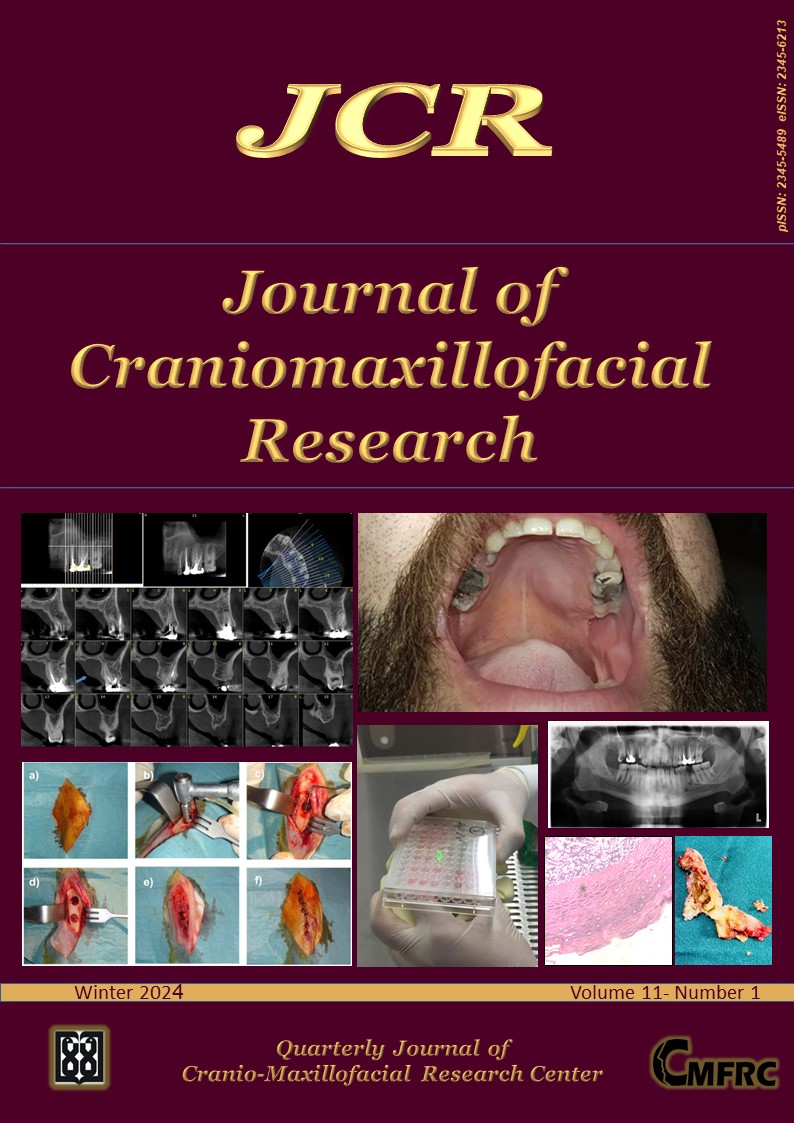
The Journal is now indexed by Scopus.
Vol 11, No 1 (Winter 2024)
Review Article(s)
-
Introduction: The focus of this review study was to examine and analyze the impact of vitamins and minerals on dental implants. Materials and Methods: In order to obtain information on the effects of vitamins and minerals on dental implants, a group of existing articles was thoroughly examined for their findings. This examination process involved the selection of specific articles that met the necessary criteria to be included in the analysis, which were then subjected to an extensive review process. The duration of this review process was over a time span of 11 years, starting from the year 2011 and concluding in the year 2022.Results: A total of 23 articles were included in the study, consisting of 9 clinical studies, 6 animal studies, 1 laboratory study, and 7 reviews. Clinical studies were limited due to uncertain results regarding the impact of vitamins and minerals on DIT, with most focusing on vitamin D levels rather than other nutrients.Conclusion: It has been concluded that vitamins and minerals play an important role in forming bone tissue. The lack of these essential elements may result in several diseases, such as diabetes mellitus and osteoporosis. It is therefore recommended to increase the levels of vitamins and minerals in patients before undergoing dental implant treatment (DIT), even though the quality of the patient’s bone can also impact the success of the implant. Consequently, the patient’s diet should be modified, and essential supplements and vitamins should be administered to ensure optimal implant success.Keywords: Vitamins; Minerals; Vitamin D; Calcium; Dental implants; Osseointegration.
Original Article(s)
-
Introduction: The aim of this study was to investigate the histomorphometric effects of bone marrow-derived mesenchymal stem cells (MSCs) transfected with Adeno Associated Virus (AAV) and containing bone morphogenic protein 7 (Bmp7) or osteoprotegerin (OPG) on bone formation after injection into inter-premaxillary suture in rats with Bmp7 or OPG alone.Materials and Methods: In 4 groups (each group n:9), different chemical solutions, namely AAV-Bmp7, AAV-OPG and AAV-Bmp7-OPG and AAV-EGF (control group) were injected into the interpremaxillary suture of rats. Bone volumes (BV), soft tissue volume (STV) and total bone volumes (TBV) of 10μm serial selection with hemotoxylin and eosin staining were calculated according to the Cavalieri principle. Each point in the region of interest was 1000μm3.Results: The BV for AAV-Bmp7, AAV-OPG, AAV-Bmp7-OPG and AAV-EGF were 46.98±1.50 mm3, 49.40±4.72mm3, 42.58±2.89mm3 and 38.82±0.76mm3, respectively. The STV was 11.53±0.99, 13.31±1.88, 8.00±4.43 and 9.57±1.90mm3 for Bmp7, OPG, AAV-Bmp7-OPG and AAV-EGF, respectively. TBV was 58.34±2.28mm3, 63.83±5.17mm3, 53.74±3.34mm3 and 48.13±1.54mm3 for AAV-Bmp7, AAV-OPG, AAV-Bmp7-OPG and AAV-EGF, respectively. The comparison between BV, STV, TBV for AAV-OPG showed a statistically significant difference (p=0.001) compared to AAV-Bmp7 or AAV-Bmp7-OPG and AAV-EGF.Conclusion: During tooth movement and bone remodeling, the ratio of soft and bone tissues is maintained by OPG. Although Bmp7 is not as effective as OPG in bone remodeling, both can reduce the retention time and the risk of recurrence. Keywords: Histomorphometry; Bmp 7; OPG; Maxillary expansion; Orthodontics; Rats.
-
Introduction: Oral squamous cell carcinoma is a multifactorial disease that is the sixth most common cancer worldwide. MicroRNAs have been confirmed to play a role in oral squamous cell carcinoma, acting as either oncogenes or tumor suppressor genes. This study examined the expression level and role of microR-148 and microR-375 in oral cancer.Materials and Methods: In this study, we used 30 cancer samples with infection and 30 cancer samples without infection. To analyze the expression of microRNA 375 and microRNA 148, we used real-time PCR. First, we extracted total RNA from the samples. Then, we generated cDNA from it. Finally, the obtained cDNA was used in the real-time PCR technique.Results: In cancer patients with oral infection, there was an increase in microRNA-148 expression and a decrease in microRNA-375 compared to cancer patients without oral infection.Conclusion: The downregulation of microRNA-375 and upregulation of microRNA-148 can be utilized as diagnostic biomarkers and prognostic factors in oral cancer.Keywords: Oral cancer; OSCC; Realtime pcr; MiR-148; MiR-375.
-
Introduction: Low-level laser therapy is a noninvasive method with the potential ability to change the balance of cell mediators and gene expressions. It affects cellular function resulting in beneficial clinical effects. This study aims to assess the effect of low-level light therapy (LLLT) using four different laser wavelengths on oral carcinoma cell viability in vitro. Materials and Methods: HN5 human head and neck squamous cell carcinoma cell lines (HNSCC) were cultured and irradiated using four wavelengths of blue (485nm), green (532nm), red (660nm), and near Infra-red (810nm) in a continuous mode with a dose of 1 J/cm2 (0.1W, 10sec) every 24hours for five consecutive days. Cell viability was assessed by evaluating mitochondrial activity by MTT assay. Results: All the wavelengths resulted in reduced viability of these cells compared to the controls. (P<0.05) There were statistically significant differences in cell viability between different wavelengths (P<0.001). The 810nm laser irradiation showed the highest percentage of cell survival (55.92%) while 660nm induced the lowest (36.02%). Conclusion: Different laser wavelengths may result in different effects on irradiated cells and red irradiation showed the lowest cell viability and the infrared laser had the highest cell viability results. Keywords: Low-level laser therapy; LLLT; Head and neck squamous cell carcinomas; HNSCC.
-
Introduction: Improving bone healing by various methods is one of the research areas of oral and maxillofacial surgery, as in the surgical branches dealing with hard tissue in medicine. Many methods, such as bone grafts, drugs, hormones, and tissue engineering applications, are used in bone reconstruction and rehabilitation. Moreover, improving bone healing is critical for better surgical outcomes. This study aimed to compare early and late period effects of intermittent teriparatide application on bone graft and local simvastatin application from histopathological, histomorphometric analysis, and biochemical aspects. Materials and Methods: 24 New Zealand rabbits were divided into four groups. While experimental groups received intermittent teriparatide (30µg/kg), control groups were given sterile distilled water. A total of 3 defects were created on each rabbit’s right and left tibia. Bone graft and local simvastatin were randomly applied to the opened defects, and one defect was left blank for control purposes. Rabbits were sacrificed on days 15 and 30 to examine early and late bone healing. Blood was drawn for biochemical analysis. Results: The healing score and new bone development in teriparatide applied and grafted defects were statistically significant compared to all groups (p<0.05). A statistically significant difference was obtained in grafted defects compared to the control group in teriparatide-applied defects. Local simvastatin caused necrosis in both experimental and control groups. Teriparatide administration does not cause a statistically significant change in calcium, potassium, and parathyroid hormone biomarkers (p>0.05).Conclusion: Better bone healing and bone graft healing in rabbits treated with teriparatide may encourage improved surgical outcomes in clinical practice. Keywords: Teriparatide; Intermittent; Bone healing; Bone graft; Simvastatin.
-
Introduction: The study aims to evaluate the oral health index related to the quality of life in patients with temporomandibular disorder (TMD) referred to the Mashhad School of Dentistry between 2018 and 2019 before and three months after treatment. Materials and Methods: This observational prospective study was conducted by interviewing patients with temporomandibular joint (TMJ) pain and clicking who referred to the Department of Prosthodontics, Mashhad Dental School, Iran, between 2018 and 2019. The demographic information of 63 patients was recorded separately. Then, using the Persian version of the OHIP-14 and GHQ-28 questionnaire, the quality of life in patients with TMD was compared before and three months after stabilization splint treatment. To evaluate the quality of life, the OHIP-sum index was used, and finally, the obtained data were analyzed by SPSS software using the Mann-Whitney test, paired-sample t-test, and Wilcoxon test.Results: OHIP sum was 24.31±8.82 and 13.15±9.52, before and after the intervention respectively. The GHQ sum was 33.32±12.9 and 20.71±11.07 before and after the intervention respectively, showing a significant decrease. The most problems of the stabilization splint users were related to psychological discomfort and the least common problems were related to functional limitations. Conclusion: The treatment of TMD using stabilization splints can significantly increase the oral health-related quality of life (OHRQoL) in these patients. Keywords: Oral health-related quality of life (OHRQoL); Temporomandibular disorders (TMD); Stabilization splint (SS).
-
Introduction: This study aimed to assess Serum Vitamin D level and female teenage caries experience. Materials and Methods: This study evaluated 330 healthy female Iranian students between 13 to 19 years residing in Rey city. A questionnaire collected their demographic information. They underwent clinical dental examination to determine the number of decayed (D), missing (M) and filled (F) teeth (DMFT index). The nutritional status was evaluated using the food frequency questionnaire (FFQ) by assessing the consumption of cariostatic, cariogenic, remineralizing, sticky foods and carbohydrates. The serum samples were collected to measure the serum vitamin D level using high-performance liquid chromatography (HPLC). Data were analyzed by multiple logistic regression and independent samples t-test. Results: The mean DMFT was 5.34±3.94. of all, 69.3% had severe vitamin D deficiency, 20.3% had moderate vitamin D deficiency and 10.4% had normal level of vitamin D. Cariostatic agents consumption had a significant inverse correlation with DMFT (P=0.006). An increase in serum level of vitamin D by more than 10.27ng/mL was associated with a reduction in prevalence of dental caries by 22%. Increased consumption of cariostatic agents by more than 89.30g/day decreased the prevalence of dental caries by 32%. No significant association was noted between the prevalence of dental caries and level of parent’s education, consanguinity of parents, level of income, place of residence, frequency of tooth brushing, dental flossing, and dental visits (P>0.05). Conclusion: While the serum vitamin D level had no significant effect on DMFT, the nutritional regimen seemed to play a more important role in caries control. Keywords: Vitamin D; Caries; Female; Teenagers; Cariostatic.
Case Report(s)
-
Salivary gland tumors are rare and account for only 2-3% of head and neck tumors, most of which are benign. Pleomorphic adenoma is the most common salivary gland tumor. This tumor mostly involves the parotid gland. However, if it occurs in the minor salivary glands, the palate is the most common site, followed by the lips, buccal mucosa, floor of the mouth, tongue, tonsils, pharynx, retromolar trigone, and gingiva. It usually presents as a slow-growing, painless submucosal mass on the hard palate. For a definitive diagnosis, it is necessary to perform a preoperative core biopsy for histopathological examination and Computed Tomography to evaluate the erosion of the hard palate and the severity of the erosion. We aim to describe the clinical, and radiological features, as well as the management of this rare localization of pleomorphic adenoma. In this case, a 30-year-old Iranian male patient with pleomorphic adenoma of the small salivary glands of the hard palate with the chief complaint of painless swelling on the left side of the palate for the past 5 years was reported. Although pleomorphic adenoma is a common entity, it is still a challenging tumor for pathologists, radiologists and surgeons. Various histological and topographic features make this tumor unique. Computed tomography and correct histopathological diagnosis are necessary to establish an appropriate surgical treatment, to achieve complete removal of the lesion through extensive local excision with periosteum or bone removal if involved to prevent recurrence.Keywords: Hard palate; Pleomorphic adenoma; Salivary glands; Rare benign tumor.
-
Epidermoid cysts are benign, rare which can occur all throughout the body of an individual. Their occurrence in the oral cavity is rare and is difficult to distinguish from other intraoral cysts. A 27-year-old female patient presented with a swelling to the oral and maxillofacial surgery department for the removal of the cystic lesion. Based on the clinical and radiographic features the provisional diagnosis was given as odontogenic keratocyst. But the histopathological examination revealed a stratified squamous cystic epithelium with abundant keratin suggestive of an epidermoid cyst. This case report presents an uncommon finding in the oral cavity with a history of teeth extraction. Based on these findings even though epidermoid cyst is rare it should be included in the differential diagnosis of radiolucent lesions of the jaws. Keywords: Sebaceous cyst; Cytokeratin; Keratohyalin granules; Teratoma; Dermoid cyst.


